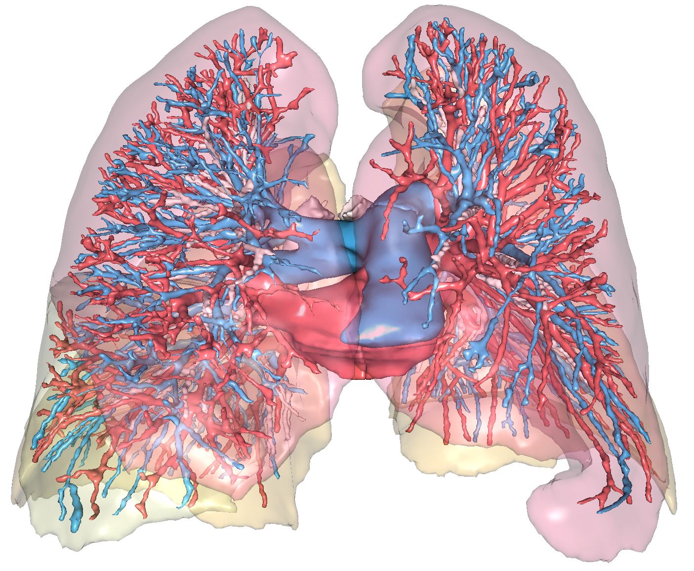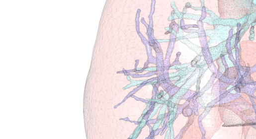
First online laboratory for 3D modelling, Visible Patient realizes from CT-scan or MRI images the 3D reconstruction of lungs.
3D lung modelling is useful to:
These 3D digital clones can be coupled with simulators to improve training, or to surgical robots to guide the gesture of surgeons.
The 3D modelling of lungs by Visible Patient can be used for following anatomical variations or pathologies:

Covid-19 context – Extensive research into the extent to which the virus affects the lungs is currently being carried out by the Visible Patient teams and the Strasbourg University Hospital, in partnership with the Nancy Hospital.
Entitled “Towards a New 3D Diagnosis of the Severity of lung infections, representative of the evolution of the patient’s state of health over 7 days“, the aim of the 3D analysis delivered by Visible Patient is to provide a rapid, quantitative and accurate measurement of the volume of the lung affected, based on a medical image.
3D lung modelling is useful for many applications:
3D organs modelled by Visible Patient can be provided in various formats, in particular STL, VTK, GLB, VPZ…
Furthermore, the 3D models can be exploited from software developed by Visible Patient that is available free of charge on PC, Mac, iOS, virtual and augmented reality head-mounted displays.

The virtual and augmented reality model visualisation software (VP Render) has not been certified as a medical device and cannot be used for clinical purposes on humans. It can nevertheless be used for training purposes as an educational, study or research tool.
Visible Patient reconstructs all the organs of the human body.
